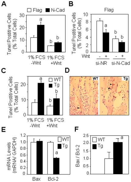
Figure 6. Forced expression of N-cadherin decreases cell survival.
(A) Control (Flag) and N-cadherin (N-Cad) overexpressing cells cultured in serum deprived (1% FCS) medium were treated with Wnt3a (15% CM) for 24 hours and the number of TUNEL-positive cells was determined. Means are +/− SD (a, P<0.05 vs -Wnt Flag cells; b, P<0.05 vs corresponding Flag cells). (B) N-cadherin silencing decreases osteoblast apoptosis. Flag cells were transfected with N-cadherin si-RNA or a non relevant si-RNA (si-NR) and treated with Wnt3a CM (15%) for 24 hours in serum deprived (1% FCS) medium and cell replication was determined. (a, P<0.05 vs -Wnt si-NR cells; b, P<0.05 vs corresponding Flag cells). (C) Osteoblasts isolated from calvaria in wild type (WT) or N-cadherin transgenic mice (Tg) were cultured in serum deprived (1% FCS) medium and treated with Wnt3a (15% CM) for 24 hours and the number of TUNEL-positive cells was determined (a, P<0.05 vs WT cells; b, P<0.05 vs corresponding WT cells. (D) Histologial sections of tibias showing increased cell apoptosis in Tg mice compared to WT mice, as revealed by TUNEL staining (brown nuclei) in osteoblasts (Ob, arrows) (x250). (E, F) Decreased Bcl-2 mRNA levels and increased Bax/Bcl-2 ratio in tibias of Tg mice compared to WT mice (a, P<0.05 vs WT mice).

