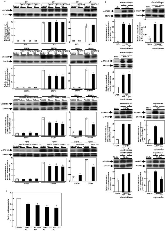
Figure 4. Signaling by specific factors was decreased in PAPST-KD cells, but not in CS chain-depleted cells.
(A) and (B) Western blot analysis of cells stimulated with the extrinsic factors. Cell lysate was prepared as described in Materials and Methods. Two independent experiments were performed and representative results are shown. The histograms show mean densitometric readings±SD of the phosphorylated protein/loading controls. Values were obtained from duplicate measurements of two independent experiments and significant values are indicated; *P<0.05, in comparison to the stimulated control; ND, not detected. (C) Luciferase reporter assay. Relative luciferase activities (TOPFLASH/FOPFLASH) are shown as means±SD from three independent experiments after normalization against the values obtained with control cells (value = 1), and significant values are indicated; *P<0.05, in comparison to the control.

