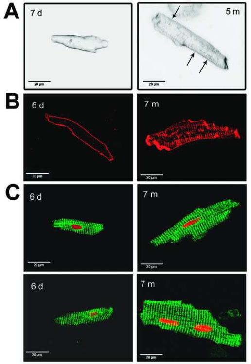
Figure 5.
Morphological analysis of ventricular cells isolated from ventricle from newborn and infant patients. A. Single ventricular cells from a newborn (left, 7 days) and an infant (right, 5 mos.) with the membrane of the cells labeled with di-8-ANNEPS. Arrows indicate partial T-tubule development in infant cell. B. Immunostaining of ventricular cells for Na/Ca exchange (NCX) in newborn (left, 6 days) and infant (right, 7 mos.). C. Immunostaining of newborn (left) and infant (right) ventricular cells with SR Ca-ATPase pump (SERCA, green) and nuclear staining (red).

