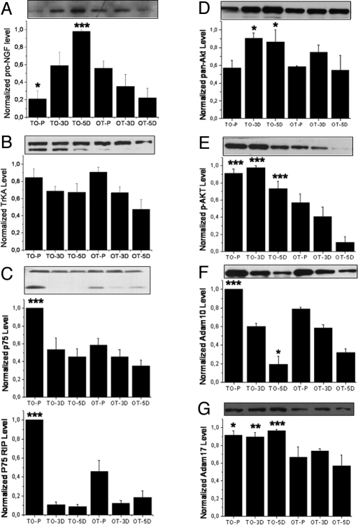
Fig. 6.
Western blots for proteins relevant to neurite outgrowth. (A) The level of proNGF increased very rapidly in TO cells upon differentiation instead of decrease, as in the case of OTcells (6A: P = 0.03, 0.302, <0.001 for P, 3D, and 5D cells, respectively). (B) For TrkA neurotrophin receptor, only the 140-kDa band was quantified because it represents the mature form of the functional NGF receptor. No difference could be detected between TO and OT cells (P = 0.612, 0.841, and 0.222 for the P, 3D, and 5D cells, respectively). However, in TO cells the 110-kDa form of the TrkA (underglycosylated immature precursor) was up-regulated. (C) The level of neurotrophin receptor p75NTR and its RIP appeared to increase dramatically in proliferating TO cells (upper band: P < 0.001, 0.443, and 0.203; lower band RIP: P < 0.001, 0.734, and 0.245, respectively for P, 3D, and 5D cells). (D and E) The level of total Akt (D) and S473 phospho-Akt (E) were in general increased in TO cells, especially the S473 phosphorylated form of Akt (for pan-Akt: P = 0.789, 0.033, and 0.042 for P, 3D, and 5D cells respectively; for p-Akt: P < 0.001, <0.0001, and <0.0001 for P, 3D, and 5D cells, respectively). (F) The level of Adam 17 increase in TO cells in all states examined (P = 0.0111, 0.002, and <0.001, for P, 3D, and 5D cells, respectively). (G) The level of Adam 10 increased in proliferating but not differentiated TO cells (P < 0.001, 0.5931, and 0.0576 for P, 3D, and 5D cells, respectively). (A–C) Typical result out of three runs performed in three repeats. Densitometric analysis as in Fig. 5. Mean and SE are indicated.

