
Figure 6.
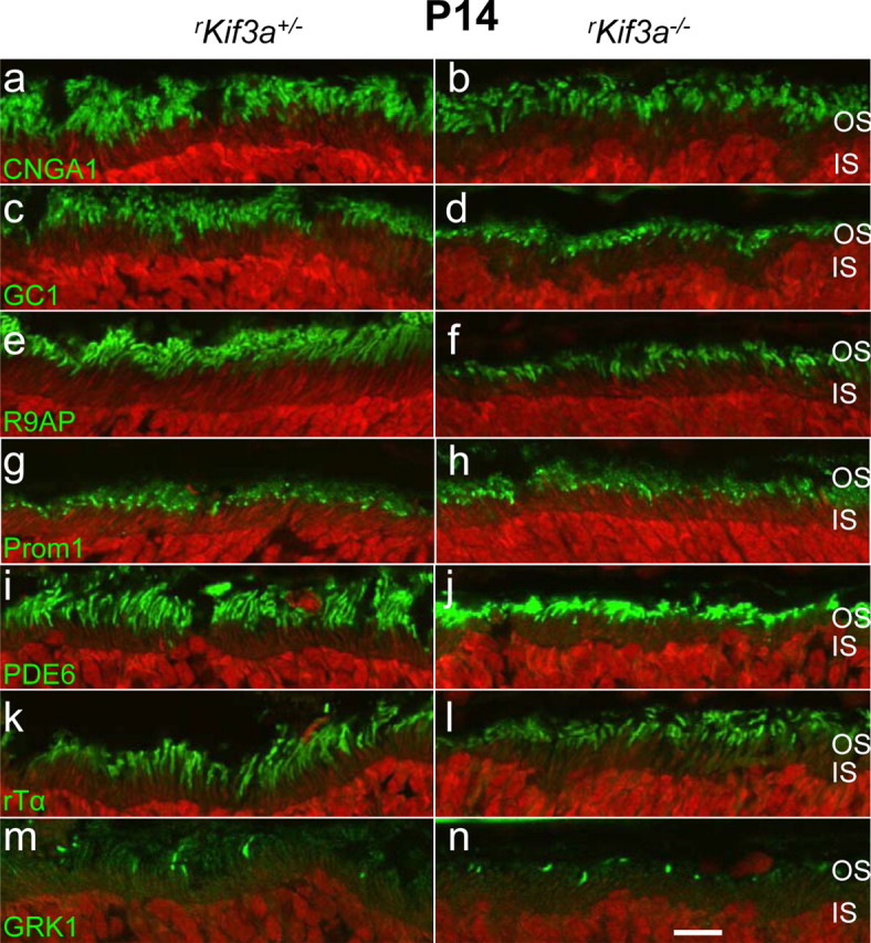
Confocal localization of phototransduction proteins. Sections of rKif3a+/− (a, c, e, g, i, k, m) and rKif3a−/− (b, d, f, h, j, l, n) retina were probed with anti-CNGA1 (a, b), anti-GC1 (c, d), anti-R9AP (e, f), anti-prom1 (g, h), anti-rod PDE6 (i, j), anti-rod Tα (k, l), and anti-GRK1 (m, n) antibodies. All panels show normal transport and localization of membrane proteins to the outer segments. Scale bar, 10 μm.