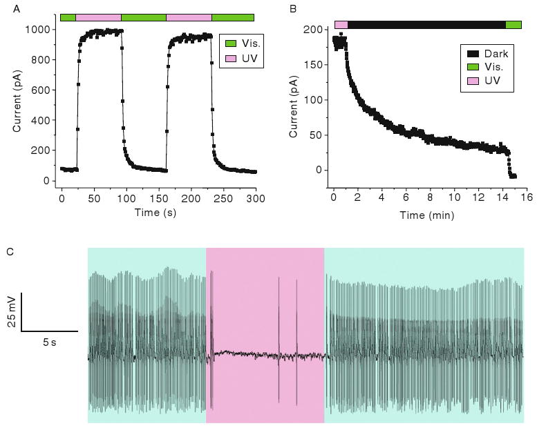
Fig. 2.

Photoswitching of SPARK channels. (A) UV light opens channels and visible light closes channels in an inside–out patch taken from a MAL-AZO-QA-treated Xenopus oocyte expressing SPARK channels. (B) SPARK channels close slowly in the dark as the photoswitches relaxes back to the trans configuration. (C) Suppression of spontaneous action potentials by UV light (380 nm) in a MAL-AZO-QA-treated cultured hippocampal neuron expressing SPARK channels. Exposure to visible light (500 nm) restores spontaneous firing.
