Figure 2.

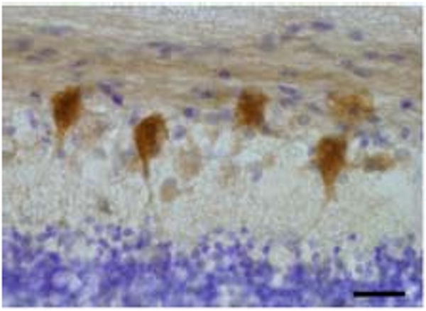
Representative high power image of human retina immunoreactive for wolframin. Retinal ganglion cells are intensely labeled for wolframin. Section is counterstained with cresyl violet. Magnification bar equivalent to 40 μm.

Representative high power image of human retina immunoreactive for wolframin. Retinal ganglion cells are intensely labeled for wolframin. Section is counterstained with cresyl violet. Magnification bar equivalent to 40 μm.