
Fig. 3.
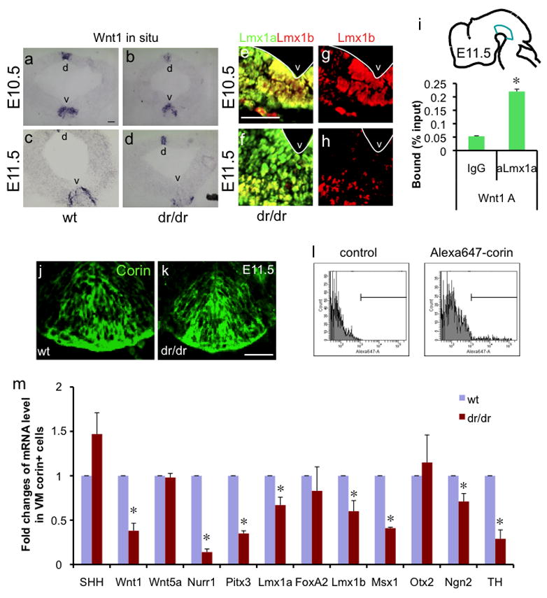
Lmx1a regulates Wnt1 expression during embryonic midbrain development. a–d. In situ hybridization analysis of Wnt1 expression. Coronal mesencephalic section of E10.5 (a, b) and E11.5 (c, d) littermate wt or dr/dr embryos. d marks dorsal mesencephalon and v marks VM. e–h. Lmx1b is expressed in the entire ventral midbrain of E10.5 embryos but is restricted to the ventral most part in E11.5 embryos. Coronal midbrain sections were stained using Lmx1b or Lmx1a antibody. The white line marks ventricle. Scale bar represents 50μm. i. E11.5 VMs were dissected as illustrated and used for ChIP using Lmx1a antibody. Binding of Lmx1a to the Wnt1 promoter was assayed by qPCR (n=3, p<0.05). j–k. Anti-corin antibody used for FACS purification marks the mDA domain, as shown in E11.5 VM of littermate wt or dr/dr embryo. Scale bar represents 50μm. l. FACS purification of mDA domain cells of littermate wt and dr/dr after staining with anti-corin antibody and Alexa-647-conjugated secondary antibody. The corin+ population is marked. m. qPCR analysis of purified mDA domain cells on the expression of regulators of mDA neuronal development. The result is the average from three independent FACS purifications (n=3, p<0.05).