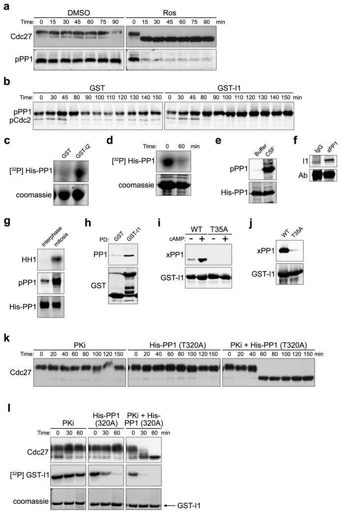
Figure 4. PP1 autodephosphorylation and I1 dephosphorylation control PP1-regulated Cdc2 substrate dephosphorylation.
A. CSF extracts treated with DMSO or Ros (0.28 mM) were immunoblotted for Cdc27 and PP1pT320.
B. GST or thio-phosphorylated GST-I1 was added to cycling extracts and samples were immunoblotted with Cdc2pY15 (mitotic entry coincides with Y15 dephosphorylation) and PP1pT320 antibodies. Full scan in S. Fig. 5F.
C. Ni-NTA agarose-bound His-PP1 was phosphorylated by Cdc2/Cyclin B in the presence of GST or GST-I2 and γ32P ATP for 30 min. His-PP1 phosphorylation was detected by SDS-PAGE/phosphorimager. Full scan in S. Fig. 5G.
D. Ni-NTA agarose-bound His-PP1 was pre-phosphorylated with high concentrations of Cdc2/Cyclin B and γ32P ATP for 30min. Ros (0.28mM) was then added and reactions were either stopped with sample buffer immediately or were incubated in buffer for an additional 60 min before resolution by SDS-PAGE/phosphorimager.
E. His-PP1 incubated with buffer or CSF extracts for 30 min, was retrieved and immunoblotted with anti-PP1pT320 or anti-His antibody. Full scan in S. Fig. 5H.
F. Protein A Sepharose-bound Xenopus PP1 antibody or control IgG were incubated with mitotic extracts for 1hr. Immunoprecipitates were blotted with anti-I1 antibody. Full scan in S. Fig. 5I.
G. His-PP1 protein was dipped into interphase or mitotic extracts for 10 min, washed and immunoblotted with anti-PP1pT320 or anti-His antibody. HH1 phosphorylation is shown.
H. GST or GST-I1 incubated with nocodazole-arrested HeLa lysates were retrieved and immunoblotted for GST or PP1.
I. Glutathione Sepharose-bound GST-I1 (WT) or GST-I1T35A incubated in interphase extracts +/− cAMP for 1 hour were washed and immunoblotted with anti-Xenopus PP1 or anti-GST antibodies. Full scan in S. Fig. 5J.
J. Glutathione Sepharose-bound GST-I1 (WT) or GST-I1T35A were incubated in mitotic extracts for 1 hour, washed, and immunoblotted with anti-Xenopus PP1 or anti-GST antibodies.
K. CSF extracts incubated with PKA specific inhibitor (PKi) H89 (0.2 mM), rabbit His-PP1T320A (2 μM) or PKI together with His-PP1T320 were immunoblotted with anti-Cdc27 antibody.
L. Glutathione Sepharose-bound GST-I1 was prephosphorylated by PKA and γ32P ATP, washed and added to CSF extracts. Extracts were treated as in 4K except GST-I1 was also retrieved and visualized by SDS-PAGE/phosphorimager.

