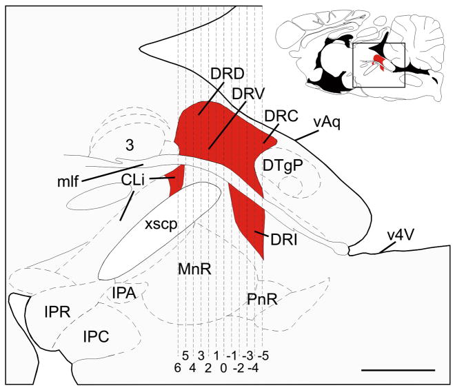
Figure 6.
Schematic illustration of a midline sagittal section of the rat brainstem showing the subdivisions of the dorsal raphe nucleus (DR) and the caudal linear nucleus (CLi) that were selected for analysis (adapted from Paxinos and Watson, 1998). Red shading indicates regions of the DR and CLi containing slc6a4 mRNA expression. The full sagittal section is shown in the top right corner for reference; the box shows the area illustrated in the main figure. Dashed vertical lines correspond to the rostrocaudal levels selected for analysis, illustrated in Figure 1. Abbreviations: 3, oculomotor nucleus; CLi, caudal linear nucleus; DRC, dorsal raphe nucleus, caudal part; DRD, dorsal raphe nucleus, dorsal part; DRI, dorsal raphe nucleus, interfascicular part; DRV, dorsal raphe nucleus, ventral part; DTgP, dorsal tegmental nucleus, pericentral part; IPA, interpeduncular nucleus, apical subnucleus; IPC, interpeduncular nucleus, caudal subnucleus; IPR, interpeduncular nucleus, rostral subnucleus; mlf, medial longitudinal fasciculus; MnR, median raphe nucleus; PnR, pontine raphe nucleus; vAq, ventral surface of the cerebral aqueduct, v4V, ventral surface of the 4th ventricle; xscp, decussation of the superior cerebellar peduncle. Scale bar, 1mm.

