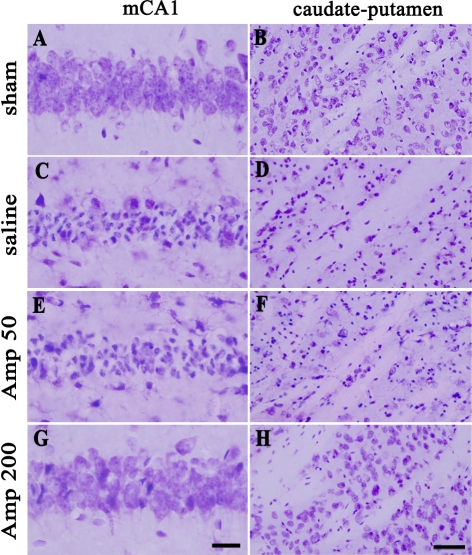
Fig. 2.
Representative photomicrographs of cresyl-violet-stained ischemic neuronal damage in the hippocampus (A, C, E, G) and the caudate-putamen (B, D, F, H) three days after transient global forebrain ischemia. sham, sham-operated group; saline, saline-treated group; Amp 50, ampicillin (50 mg/kg/day)-treated group; Amp 200, ampicillin (200 mg/kg/day)-treated group. Scale bar=20 µm (A, C, E, G), 50 µm (B, D, F, H).

