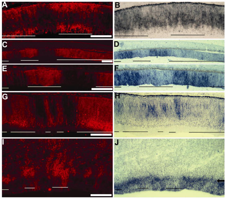
Fig. 4.
Diminished ash1 expression in E7.5 retinas infected by RCAS-ngn1. A, B: Control RCAS-GFP retina with double-labeling for RCAS viral protein p27 (A) and for ash1 mRNA (B). C–H: RCAS-ngn1 partially-infected retinas showing double-labeling for RCAS viral protein p27 (C, E, G) and for ash1 mRNA (D, F, H) in peripheral retina (C–F) and central retina (G, H). C and D are lower magnifications of E and F. I, J: Double-labeling for viral protein p27 (I) and ash1 expression (J) of E7.5 brain partially infected with RCAS-ngn1. Arrow in J points to the zone with diminished ash1 expression. In all panels, infected regions are approximately underlined. Scale bars: 100 μm.

