
Figure 5. P68 nucleocytoplasm shuttle is RanGTPase pathway dependent.
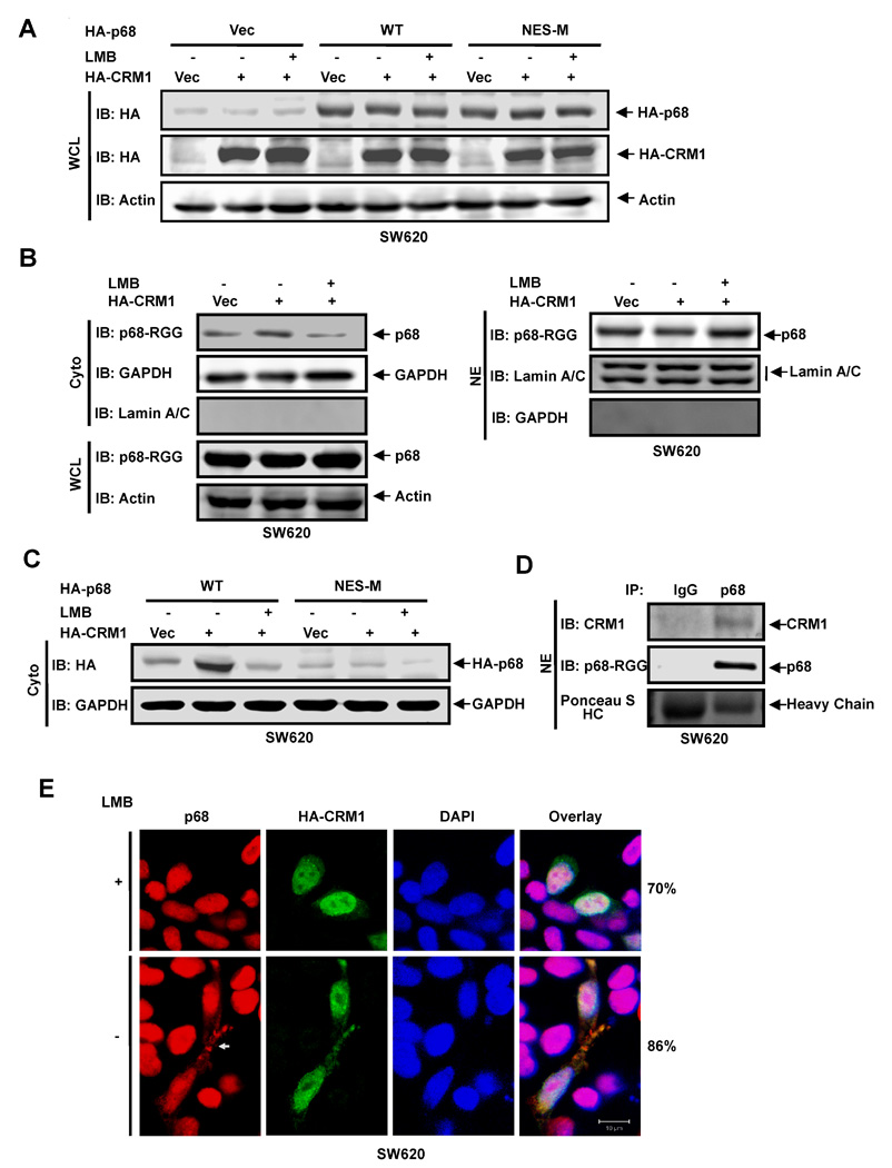
(A) Exogenous expression of HA-p68s, wild-type (WT) and mutant (NES-M), and HA-CRM1 in SW620 cells were analyzed by immunoblot of whole cell lysate (WCL) using anti-HA antibody (IB:HA). (B) (Left panel) Cytoplasmic (Cyto) p68 levels of SW620 cells were analyzed by immunoblot using the antibody p68-rgg (IB:p68-RGG). Immunoblots of p68 in whole cell lysate (IB:p68-RGG) indicate cellular levels of p68. (Right panel) Nuclear (NE) levels of p68 in SW620 cells were analyzed by immunoblot using the antibody p68-rgg (IB:p68-RGG). (C) The cytoplasmic (Cyto) levels of exogenously expressed HA-p68s (WT and NES-M) were analyzed by immunoblot of cytoplasmic extracts using anti-HA antibody (IB:HA). In (A), (B), and (C), IBs of β-actin (IB:actin), GAPDH (IB:GAPDH), Lamin A/C (IB:Lamin A/C) are controls. The SW620 cells were transfected with vector alone (Vec), the vector for CRM1 (HA-CRM1), and the vector for CRM1 plus treatment with LMB (HA-CRM1 + LMB). (D) Co-immunoprecipitation of p68 with endogenous CRM1 in nuclear extracts (NE) of SW620 cells was analyzed by immunoprecipitation using the antibody Pabp68 (IP, p68). The immunoprecipitates were examined by immunoblot using antibody against CRM1 (IB:CRM1) and the antibody p68-RGG (IB:p68-RGG). Ponceau S staining the IgG haeve chain is the loading control (Ponceau S HC). Immunoprecipitation using rabbit IgG (IP, IgG) is negative control for co-immunoprecipitation. (E) Examples of confocal fluorescent microscopy images of SW620 cells show the cellular localizations of endogenous p68 (P68) and exogenously expressed HA-CRM1 (HA-CRM1). The cells were treated (+) or untreated (−) with LMB. Red signals are staining p68. The green signal represents staining of HA-tag. The blue signal represents staining of DNA. Overlay images are overlay of staining of p68, HA-CRM1, and DNA. The numbers are percentage of cells showing similar image pattern in a random group of 50 cells.