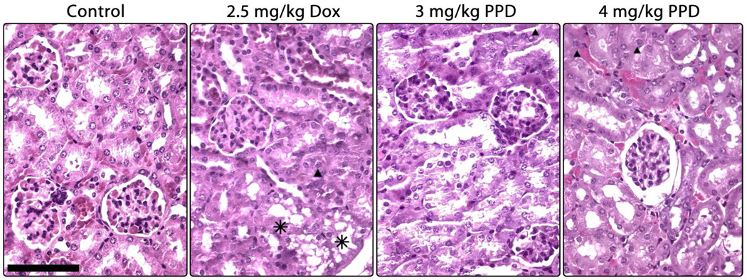
Figure 7.
Micrographs of hematoxylin/eosin stained kidney sections as a function of treatment as described in Figure 3. Tissue from control mice is healthy, exhibiting well-formed tubules, glomeruli, and cells. Treatment with 2.5 mg/kg Dox resulted in the appearance of a vacuolar-like damage (asterisks) and hyaline deposits in a subset of tubules (triangle). Neither prodrug treatment resulted in visible cellular damage, although a dose-dependent congestion of tubules with hyaline deposits (triangles) was evident. Additionally, the higher dose of PPD induced a thickening of Bowman's capsule in a small population of glomeruli, as shown on the right. The bar indicates 200 µm.

