FIGURE 7.
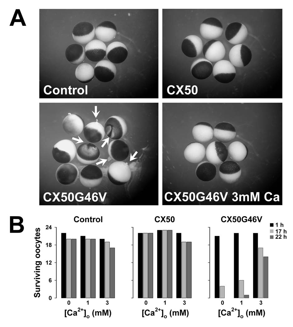
Expression of CX50G46V induced hemichannel activity-dependent oocyte death. Oocytes were injected with similar amounts of CX50 or CX50G46V cRNA and incubated in modified Barth’s solution containing 0, 1 or 3 mM Ca2+ overnight at 18°C. (A) Oocytes injected with CX50G46V cRNA showed obvious blebbing and discoloration (arrows) when incubated in modified Barth’s solution containing 1 mM Ca2+ for 17 hours (lower left panel). In contrast, oocytes injected with CX50 cRNA or oligonucleotide antisense to the endogenous Xenopus CX38 (Control) showed no apparent detrimental changes when studied under identical conditions (upper panels). The rate of cell death of the CX50G46V cRNA-injected oocytes was significantly reduced by increasing the external calcium concentration from 1 to 3 mM (lower right panel). (B) Graphs represent the number of surviving oocytes at different times following injection of cRNA in media containing 0, 1 or 3 mM Ca2+. Oocytes were scored for cell viability based on appearance as illustrated in A.

