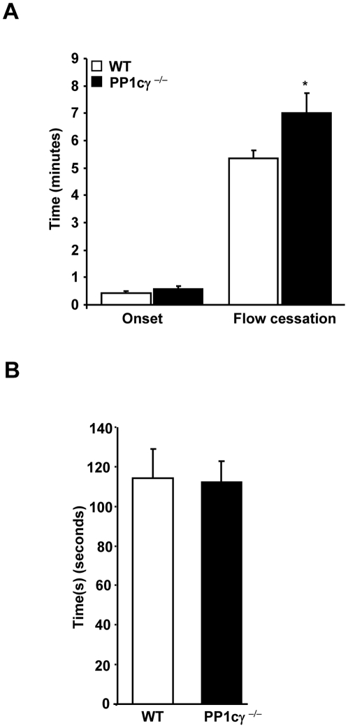
Figure 5. Role of PP1cγ in thrombosis and hemostasis injury models.
(A). Light/dye -induced microvascular thrombosis was studied in the venules of the cremaster muscles of WT and PP1cγ−/− mice by intra vital microscopy. The onset of thrombosis (onset) and the cessation of blood flow following the injury were monitored. Results are expressed as ±SEM of 13 experiments. Compared to the WT, the delayed time for flow cessation in PP1cγ−/− mice was significant at *p = 0.03. (B). Tail vein bleeding times from 13 WT and PP1cγ−/− mice are shown.

