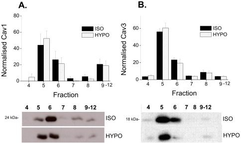
Figure 4. Hyposmotic swelling does not cause translocation of Cav 1 or Cav 3 from caveolar membrane fractions.
Ventricular myocytes were exposed to isotonic or hypotonic solutions for 15 min, then subject to Na2CO3 extraction and discontinuous sucrose density gradient fractionation. A. Mean data showing Cav 1 in each fraction normalised to the sum of Cav 1 in all fractions for each sample (n = 6 hearts). Representative immunoblots, with equal volume loading of fractions, is shown below. B. Mean data showing Cav 3 in each fraction with representative immunoblots below. In both isotonic and hypotonic conditions, Cav 1 and 3 were enriched in the buoyant lipid raft fractions (5,6) of the myocyte. Hyposmotic swelling had no effect on the location of either Cav isoform.

