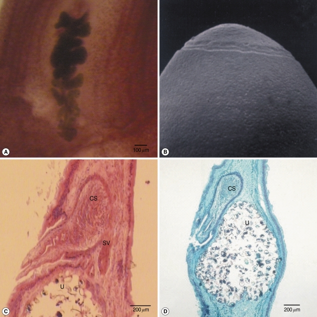
Fig. 1.
Gravid proglottids and an egg of Diphyllobothrium nihonkaiense. (A) Whole mounted specimens of a proglottid showing the uterus and cirrus sac (× 25), (B) A SEM photo of the eggshell showing shallow pits and opercular structure (× 3,000), (C, D) Longitudinal sections of a mature proglottid showing the cirrus sac (CS), seminal vesicle (SV), and uterus (U) (C: H-E stain, D: trichrome stain).

