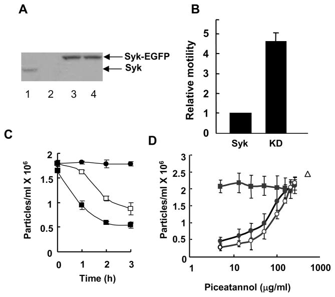
Figure 1.
Syk alters cell aggregation and motility. A, lysates from MCF10A (lane1), Syk-deficient MCF7 (lane 2) or MCF7 cells stably expressing Syk-EGFP (lane 3) or Syk-EGFP(K396R) (lane 4) were analyzed by Western blotting with an antibody against Syk (N19). Arrows mark the migration positions of the Syk-EGFP and Syk-EGFP(K396R) fusion proteins (upper arrow) and endogenous Syk (lower arrow). B, MCF7 cells stably expressing Syk-EGFP (Syk) or Syk-EGFP(K396R) (KD) were analyzed in the transwell migration assay. The cells at the lower surface of the filter were stained with DAPI and observed using fluorescence microscopy. Motility was normalized to that of cells expressing Syk-EGFP. The data indicate the mean and standard deviations of three independent experiments (P < 0.01). C, stable MCF7 cell lines expressing Syk-EGFP (■) or Syk-EGFP(K396R) (□) were allowed to aggregate for the indicated periods of time. MCF7 cells did not aggregate in the presence of EGTA (●). The number of particles formed in four individual samples was counted. The averages and standard deviations for three experiments are shown. D, MCF10A cells were allowed to form cell-cell contacts for a period of 0 (■), 1 (●) or 2 (○) h in the presence of increasing concentrations of piceatannol. The number of particles formed in 2 h in the absence of piceatannol, but in the presence of EGTA is indicated (△). The number of particles is defined as a single cell or a single cluster of cells. The data indicate the mean and standard deviations of three independent experiments.

