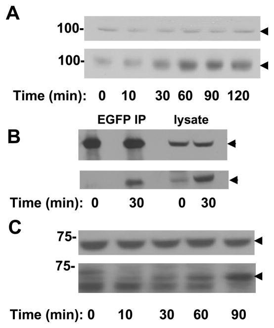
Figure 7.
Syk is phosphorylated following integrin-crosslinking. A, integrins on MCF7 cells expressing Syk-EGFP in suspension were crosslinked with anti-β1 integrin antibodies for the times indicated. Cell lysates were analyzed by Western blotting with antibodies against Syk (upper panel) or phosphotyrosine (lower panel). The migration position of Syk-EGFP is indicated by the arrowheads. B, lysate and anti-GFP immune complexes from MCF7 cells expressing Syk-EGFP and stimulated in suspension with anti-β1 integrin antibodies for the indicated times were analyzed by Western blotting with antibodies against Syk (upper panel) or phosphotyrosine (lower panel). The migration position of Syk-EGFP is indicated by the arrowheads. C, integrins on MCF10A cells in suspension were crosslinked with anti-β1 integrin antibodies for the times indicated. Cell lysates were analyzed by Western blotting with antibodies against Syk (upper panel) or phosphotyrosine (lower panel). The migration position of Syk is indicated by the arrowheads.

