Figure 8.
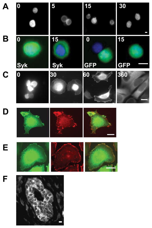
Integrin ligation alters the subcellular localization of Syk. A, MCF7 cells transfected with a Syk-EGFP-expression plasmid were stimulated by crosslinking β1 integrins for 0, 5, 15 or 30 min, fixed and examined by fluorescence microscopy. B, MCF7 cells transfected with plasmids expressing Syk-EGFP (Syk) or EGFP (GFP) were treated with β1 integrin antibodies for 0 or 15 min, fixed, stained with DAPI and examined by fluorescence microscopy. Merged images illustrating the location of Syk-EGFP or EGFP (green) and DAPI-stained nuclei (blue) are shown. C, Tet-inducible MCF7 cells treated with tetracycline to induce Syk-EGFP were plated on fibronectin-coated coverslips for the times indicated (in minutes) and then fixed and examined by fluorescence microscopy. D and E, MCF7 cells transfected with a Syk-EGFP-expression vector were plated on coverslips coated with fibronectin, allowed to spread for 60 min and then fixed, permeabilized and stained with monoclonal antibodies against vinculin (C) or with rhodamine phalloidin (D). The localization of Syk-EGFP (green, D, E), endogenous vinculin (red, D), and F-actin (red, E) were visualized with fluorescence microscopy. F, a human mammary tissue slice was stained with anti-Syk antibodies for immunohistochemical analysis. Bars = 10 μm.

