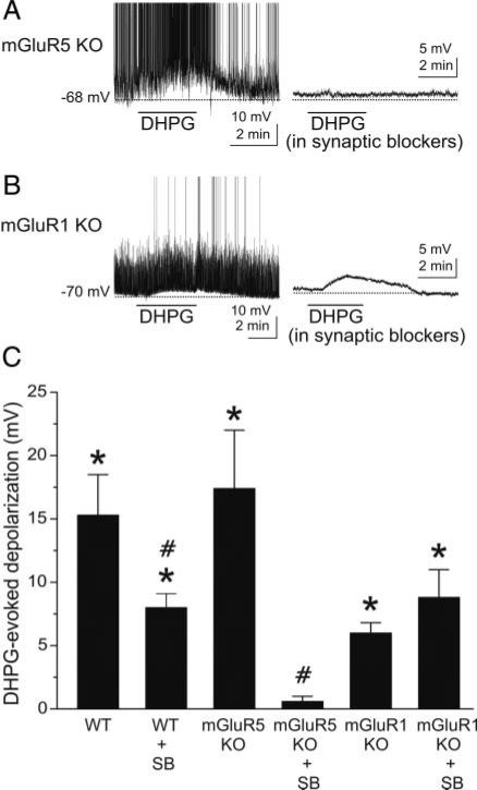
Fig. 4.
DHPG-evoked excitation of GCs is absent in mGluR5 null mutant mice. A, left: DHPG (50 μM) excites a GC recorded in a slice harvested from an mGluR5–/– mouse. Right: in the presence of synaptic blockers [CNQX (10 μM), APV (50 μM), and gabazine (5 μM)], DHPG had no effect on the same GC. B: in normal media (left), DHPG depolarizes and moderately increases the firing rate of a GC from an mGluR1–/– mouse. Subsequent application of DHPG (50 μM) to the same cell in the presence of synaptic blockers (right) elicited a comparable depolarization although evoked spikes are absent. Note that application of synaptic blockers (right)in A and B decreases baseline noise compared with left control traces, which include spontaneous EPSPs. C: bar graph showing group data on the effects of DHPG in wild-type and mGluR KO mice with or without synaptic blockers present; data are expressed as DHPG-evoked depolarization (mV) compared with control. WT, wild-type mice; mGluR5 KO, mGluR5 knockout mice; mGluR1 KO, mGluR1 knockout mice; SB, synaptic blockers; see text for numbers of GCs tested. *, significantly different from control, P < 0.05, paired t-test. #, significantly different from respective non-SB group, P < 0.05, independent t-test. See text for specific P values. The DHPG-evoked depolarization in the mGluR1 KO group was significantly less than in the WT group (P < 0.02). There were no significant differences (P > 0.05, see text for specific P values) between the 1) WT and mGluR5 KO groups, 2) WT + SB and mGluR1 KO + SB groups, and 3) mGluR1 KO and mGluR1 KO + SB groups.

