
Figure 1.
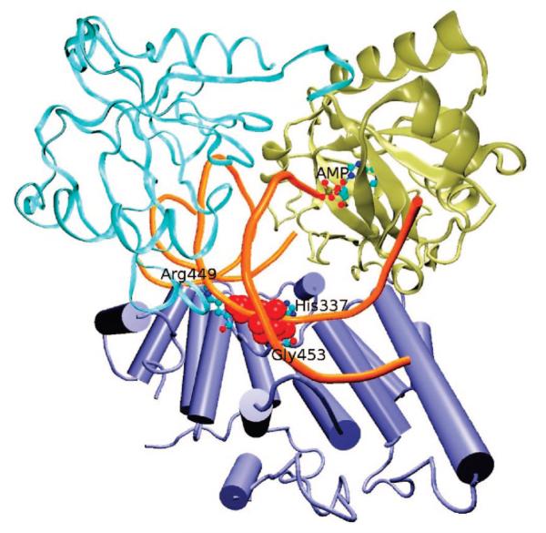
The DNA substrate (orange tube) is encircled by three domains of human DNA ligase I, i.e., the DNA binding domain (DBD) containing residues Asp262—Ser535 (ice-blue carton), the adenylation domain (AdD) Pro536—Asp748 (wide tan ribbon), and the OB-fold domain (OBD) Tyr749—Ser901 (narrow cyan ribbon). The AMP cofactor (in CPK representation) is located in AdD. The putative binding site on DBD is represented by red spheres, and the three residues defining the binding pocket, His337, Arg449 and Gly453, are shown in CPK representation.