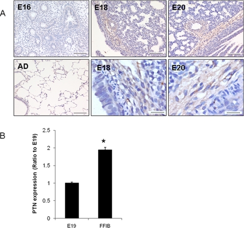
FIGURE 1.
PTN expression during fetal lung development. A, immunostaining of PTN in the developing lungs. Embryonic days 16, 18, and 20 (E16, E18, E20) and adult (AD) lung tissue sections were stained using anti-PTN antibodies and the Vectastain ABC kit. Scale, 100 μm. The lower right two panels are high magnifications (scale bar, 20 μm). B, real-time PCR analysis of PTN mRNA expression in E19 fetal lung tissue (E19) and isolated E19 fetal fibroblasts (FFIB). The results were normalized to 18 S rRNA and expressed as a ratio of lung tissue. Data shown are the means ± S.E.; *, p < 0.05 versus lung tissue (n = 3 cell preparations, assayed in duplicate).

