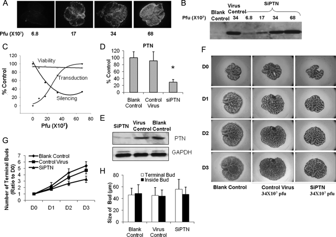
FIGURE 6.
Effect of PTN on fetal lung branching morphogenesis. A–C, E14 fetal lungs were isolated and treated with various doses of a PTN siRNA adenovirus (SIPTN) or a virus control. GFP fluorescence was taken at day 3 (A), and fetal lung tissues were analyzed by Western blot using anti-GFP antibodies (B). Pfu, plaque-forming units. The transduction efficiency, silencing efficiency, and cell viability were monitored and expressed as a percentage of untreated lungs or a maximal value (C). Transduction efficiency was measured by determining GFP expression levels. Silencing efficiency was measured by determining ptn mRNA levels using real-time PCR. mRNA levels were normalized to 18 S rRNA. Viability was measured by determining lactate dehydrogenase release. D–H, E14 fetal lungs were treated with a PTN siRNA adenovirus or a virus control (34 × 107 plaque-forming units) for 3 days. PTN mRNA (D) and protein (E) levels were determined by real-time PCR and Western blot. Photographs were taken at day 0 to day 3 (D0–D3) (F). Terminal bud numbers (G) were counted and normalized to day 0. The sizes of terminal buds on surface and inside of the lungs were also measured at day 3 (H). Data shown are the means ± S.E.; n = 6 from 3 different pregnant rats.

