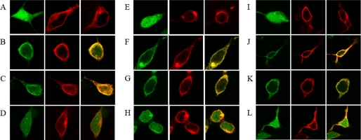
FIGURE 3.
Membrane colocalization of CaM with A2A or D2 receptors. Co-transfected HEK-293 cells were washed and resuspended for 2.5 h in HBSS buffer containing 1.26 mm CaCl2 (A–D and I–L) or in a Ca+2-free HBSS buffer containing 1 mm EDTA (E–H). A, E, and I, confocal microscopy images of HEK-293T cells expressing (from left to right in each panel) CaMYFP (0.6 μg of cDNA), A2ARRluc (1 μg of cDNA), or D2RRluc (1 μg of cDNA). B–D, F–H, and J–L, confocal microscopy images of HEK-293T cells co-transfected with the above described amounts of cDNA for CaMYFP and A2ARRluc (B, F, and J), CaMYFP and D2RRluc (C, G, and K), or CaMYFP and A1RRluc (1 μg of cDNA) (D, H, and L). Ionomycin-treated cells (10 min prior to fixation) are shown in I–L. CaM was identified by YFP fluorescence (green images), and receptor-Rluc constructs were identified by immunocytochemistry (using a monoclonal anti-Rluc primary antibody and a cyanine-3-conjugated secondary antibody). Colocalization (yellow) is shown in the right panels.

