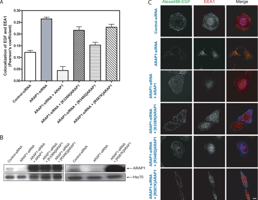
FIGURE 12.
Effect of ARAP1 on EGF traffic to EEA1-positive endosomes. HeLa cells were transfected either with control-siRNA or ARAP1-specific siRNA. After 48 h, cells were transfected with plasmids directing the expression of FLAG-ARAP1, [K336N]ARAP1, [R348Q]ARAP1, and [R587K]ARAP1. Twenty-four h later, cells were serum-starved overnight and incubated with 1 μg/ml fluorescent Alexa488-EGF for 10 min at 37 °C. The cells were fixed and immunostained with mouse anti-EEA1 (red) and rabbit anti-FLAG (blue). Pearson's coefficients for signals from EGF and EEA1 were determined for 25 cells captured with a Zeiss LSM510-Meta laser-scanning confocal microscope with a ×63, 1.4 NA Plan Neofluar oil immersion lens under each condition using Zeiss LSM 5 image software. The average and range from two experiments is shown in A. An immunoblot to determine ARAP1 levels in lysates from cells treated with siRNAs or expression plasmids, as indicated, is shown in B. Antiserum (1153) raised against ARAP1 was used to determine the ARAP1 levels, and anti-Hsc70 antibody was used as a protein loading control. Representative micrographs are shown in C. Scale bar, 10 μm.

