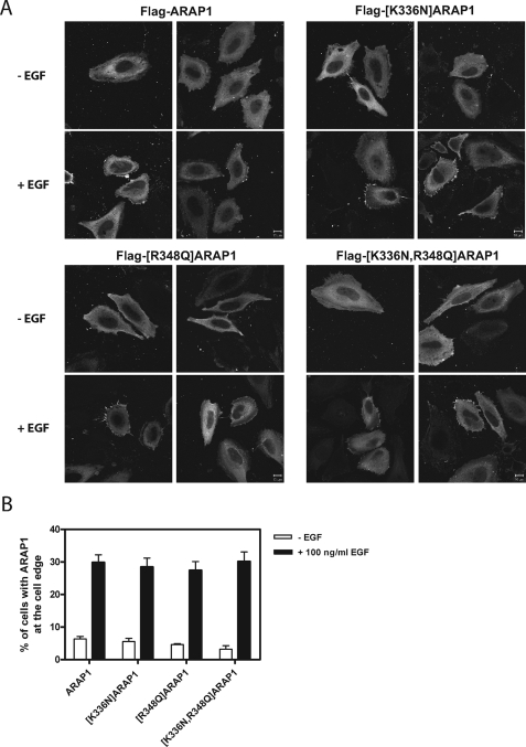
FIGURE 9.
ARAP1 association with the cell edge. A, representative images. HeLa cells overexpressing N-terminal FLAG-tagged ARAP1 or mutant ARAP1 were serum-starved for 6 h on fibronectin-coated coverslips (10 μg/ml) and treated with 100 ng/ml EGF for 5 min at 37 °C. Fixed cells were immunostained with monoclonal anti-FLAG antibody. B, quantification of localization. More than 100 cells were scored for ARAP1 concentrated at the cell edge using visual inspection in three independent experiments. Images were captured with a Zeiss LSM510-Meta laser-scanning confocal microscope with a ×63, 1.4 NA Plan Neofluar oil immersion lens. Scale bar, 10 μm.

