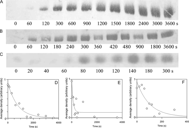
FIGURE 3.
Thr305/306 autophosphorylation time course estimated by Western blotting. 1 μm αCaMKII was allowed to autophosphorylate for 15 s, as described under “Materials and Methods.” At that point, 4.2 mm EGTA was added, and aliquots were taken at the indicated time points; the reaction was quenched by pipetting the aliquots taken into SDS sample buffer. A rabbit polyclonal anti-phospho-αCaMKII (Thr305) (Millipore) was used. Shown are Western blots and respective densitometric analyses of representative time courses at 21 (A and D), 30 (B and E), and 37 °C (C and F). Densitometric analysis of Western blots was carried out as described under “Materials and Methods.”

