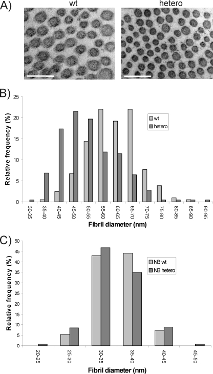
FIGURE 1.
A, transmission electron microscopy analysis of collagen fibrils in skin of adult heterozygous LH3 knock-out (hetero) and wild-type (wt) mice. Scale bar, 200 nm. B and C, relative frequencies (%) of fibril diameters in the skin of adult (hetero) and newborn (NB hetero) heterozygous LH3 knock-out mice that differed from wild-type mice (wt or NB wt). The diameters of the collagen fibril cross-sections were measured from digital images using image analysis software.

