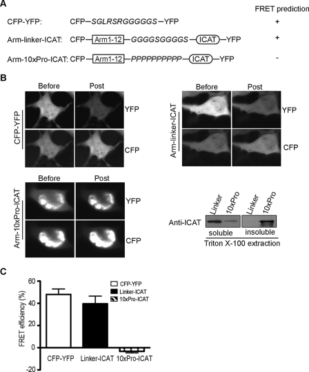
FIGURE 3.
FRET analysis of β-catenin structure in cells. A, schematic of control constructs for single molecule acceptor photobleaching FRET. A construct containing CFP and YFP fluorescent proteins separated by 12 amino acids was designed to optimize acceptor photobleaching FRET conditions. ICAT protein was linked to the β-catenin arm-repeat region by using either a flexible linker (GGGGSGGGGS) or a rigid peptide consisting of 10 proline residues to serve as positive and negative controls, respectively. B, representative FRET images and immunoblot of CFP-YFP, arm-linker-ICAT, and arm-10Pro-ICAT. Cos-7 cells were solubilized in 1% Triton X-100 lysis buffer after imaging, and the relative solubility of arm-linker-ICAT and arm-10Pro-ICAT proteins was assessed by immunoblotting using antibody for ICAT. C, FRET efficiencies for CFP-YFP, Linker-ICAT, and 10xPro-ICAT were calculated from at least 100 cells imaged from multiple transfections and are expressed as the mean ± S.D.

