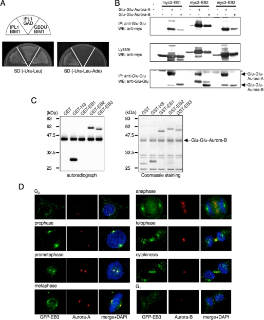
FIGURE 1.
EB2 and EB3, but not EB1, are substrates of Aurora. A, yeast Aurora homolog, Ipl1, binds Bim1, the sole budding yeast member of the human EB1-related family, in a yeast two-hybrid system. Plasmids transformed into PJ69-4A are indicated in each sector. B, binding of EB1 family members and Aurora family members in COS-7 cells that were transfected with the indicated plasmids. The cell lysates were immunoprecipitated (IP) by using anti-Glu-Glu antibody, and the precipitates were analyzed by Western blots (WB) that were probed sequentially with anti-Myc and anti-Glu-Glu antibodies. Asterisk denotes IgG heavy chain. One Western blot shown is representative of three independent experiments. C, in vitro phosphorylation of the EB1 family proteins. GST-fused EB1, EB2, EB3 or H3-(5–15) as a control was incubated with immunoprecipitated Glu-Glu-Aurora-B in the presence of [γ-32P]ATP. The phosphorylation reactions were detected by autoradiography (left panel). The amounts of GST fusion protein used in the assay were compared by Coomassie Blue staining (right panel). A representative result of three independent experiments is shown. D, representative confocal images of EB3 localization during each stage of mitosis by immunofluorescence staining of HeLa cells that stably expressed GFP-EB3. Green, GFP-EB3; red, Aurora-A (left panels) or Aurora-B (right panels); blue, DAPI.

