FIGURE 2.
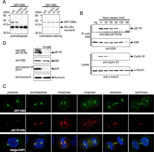
Phosphorylation of EB3 Ser-176 by Aurora-A and Aurora-B during mitosis. A, in vitro phosphorylation of EB3 mutants. The experiment was performed as described in Fig. 1C using GST-fused EB3 mutants and immunoprecipitated Glu-Glu-Aurora-B. The autoradiographs shown are representative of three independent experiments. WT, wild type. B, phosphorylation of endogenous EB3 in mitotic cells. HeLa cells were released from a nocodazole block for the indicated times. Noco and log indicate nocodazole and logarithmic phase, respectively. Immunoprecipitation (IP) was performed with anti-EB3 monoclonal antibody, and the precipitates were analyzed by Western blots probed sequentially with anti-phospho-EB3(Ser-176) rabbit polyclonal antibody (Rb). A representative result of three independent experiments is shown. C, representative confocal images of localization of phospho-EB3(Ser-176) during each stage of mitosis. Immunofluorescence staining of HeLa cells that stably expressed GFP-EB3 was performed using anti-phospho-EB3(Ser-176) mouse monoclonal antibody (mAb). Red, phospho-EB3(Ser-176); green, GFP-EB3; blue, DAPI. D, effects of Aurora inhibitor on EB3 phosphorylation. HeLa cells were arrested at prometaphase with nocodazole and subsequently treated with VX-680 for 4 h, and then the cell lysates were analyzed by Western blots (antibodies indicated at the side of the figure). A representative result of three independent experiments is shown.

