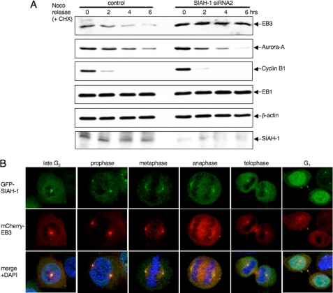
FIGURE 5.
Degradation of EB3 by SIAH-1 at the mitosis to G1 transition. A, effects of SIAH-1 knockdown on EB3 stability during the mitosis-to-G1 transition. HeLa cells were transfected with siRNA for SIAH-1. After 48 h, nocodazole was added for 16 h; subsequently, cells were released from a nocodazole (Noco) block for the indicated times in the presence of 10 μg/ml CHX. The cell lysates were subjected to Western blots with the indicated antibodies. One Western blot shown is representative of three independent experiments. B, representative confocal images of localization of SIAH-1 and EB3 during each stage of mitosis. Immunofluorescence staining is shown of HeLa cells that stably expressed GFP-SIAH-1 and mCherry-EB3. Green, GFP-SIAH-1; red, mCherry-EB3; blue, DAPI.

