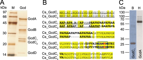
FIGURE 2.
A, silver-stained 15% SDS-PAGE analysis of the purified glutaconyl-CoA decarboxylase from C. symbiosum. Lane Gcd contains 30 μg of purified enzyme (lane M: molecular mass marker). B, comparison of the amino acid sequences of GcdC1 and GcdC2 from C. symbiosum. The N-terminal domain was sequenced by Edman degradation (underlined). Conserved residues are marked in yellow. Bold amino acids indicate the AP-rich linker region and the biotin-binding site. The highly conserved motif is highlighted by a blue box while the lysine to which biotin is attached is shown in red. C, Western blot analysis of a Ni-NTA purification of the co-expression of GcdA and biotin-containing GcdC1. The proteins were detected with avidin-peroxidase (lane B) or with anti-Penta-His-HRP conjugate (lane H). Each lane contains 5 μg of protein.

