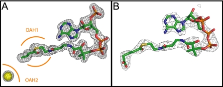
FIGURE 3.
A, final 2Fo-Fc electron density corresponding to GcdA-bound crotonyl-CoA contoured at 3 ς (1.75 Å resolution). Additional electron density found in the vicinity of the supposed biotin binding site was assigned to a chloride ion (yellow sphere). The positions of the oxyanion holes OAH1 and OAH2 are indicated in orange. B, Fo-Fc map of glutaryl-CoA contoured at 1 ς (2.7 Å resolution). This figure and Figs. 4–7 were prepared by PyMOL (58).

