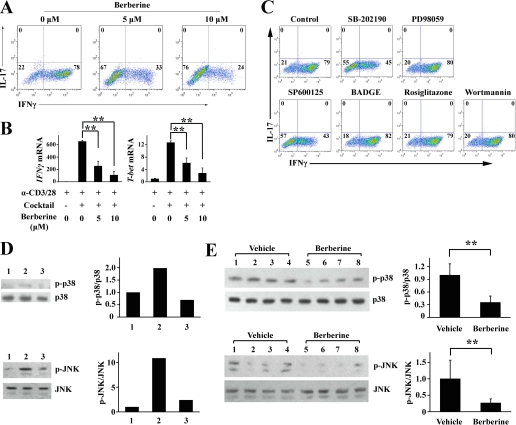
FIGURE 6.
Berberine suppress Th1 differentiation in vitro. A, naive CD4+ T cells from NOD mice were cultured under the Th1 condition in the absence or presence of berberine at the indicated concentration for 4 days. Intracellular staining profiles of IL-17 and IFNγ are shown. B, naive CD4+ T cells were isolated from NOD mice and cultured with α-CD3/28 to stimulate TCR in the absence or presence of Th1 differentiation mixture (IL-12 and α-IL-4). Berberine was then added at the indicated concentration. Three days later, RNA was extracted and reverse-transcribed into cDNA and subjected to real time PCR. Gene expression is presented relative to that of β-actin (**, p < 0.01, n = 3). C, naive CD4+ T cells from NOD mice were cultured under the Th1 condition in the presence of the indicated drugs for 4 days. Intracellular staining profiles of IL-17 and IFNγ are shown. D, naive CD4+ T cells from NOD mice were cultured in the presence of α-CD3/28 alone (lane 1), with the Th1 mixture alone (lane 2), or with the Th1 mixture plus berberine (lane 3) for 24 h. Cell lysates was subjected to Western blotting for p38 MAPK, JNK, phosphorylated p38 MAPK and phosphorylated JNK. Quantification of the optical density of the bands is shown in the right panel. E, CD4+ T cells were purified from vehicle- or berberine-treated mice, and cell lysates were subjected to Western blotting for p38 MAPK, JNK, phosphorylated p38 MAPK, and phosphorylated JNK. Quantification of the optical density of the bands is shown in the right panel (**, p < 0.01, n = 4).

