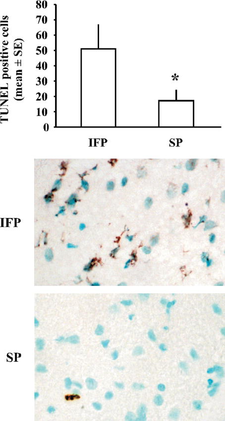
Figure 1.
TUNEL staining demonstrates decreased DNA fragmentation 22.5hr following tMCAO in rats fed a high soy diet (SP) compared to control ovariectomized female rats (IFP). Bars represent mean +/− SE of TUNEL-positive cells in the ischemic cortex (n=4/ group, *p< 0.05). Shown are representative images of TUNEL staining in the ischemic cortex of IFP and SP rats. TUNEL-positive cells are stained brown, while other cells are counterstained with methyl green.

