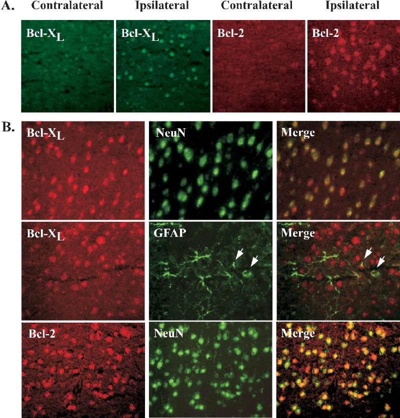
Figure 6.
IHC analysis of bcl-xL and bcl-2 22.5hr following tMCAO. (A) Representative pictures showing differential expression of bcl-xL and bcl-2 protein in the cerebral cortex of IFP rats after cerebral ischemia. In response to injury, both bcl-2 and bcl-xL expression was upregulated in the ipsilateral (ischemic) cortex of all groups compared to the contralateral (non-stroked) side. (B) Localization of bcl-xL and bcl-2 to neurons is shown in representative pictures in which bcl was double-labeled with the neuronal marker NeuN in the ischemic cortex 22.5hr following tMCAO. Overlayed images demonstrate predominant neuronal expression of both bcl-xL and bcl-2. A panel showing bcl-xL double-labeled with the astrocyte marker GFAP is included to demonstrate that bcl-xL is not found in these cells. However, arrows show close association of bcl-xL and GFAP-expressing cells.

