
Fig. 6.
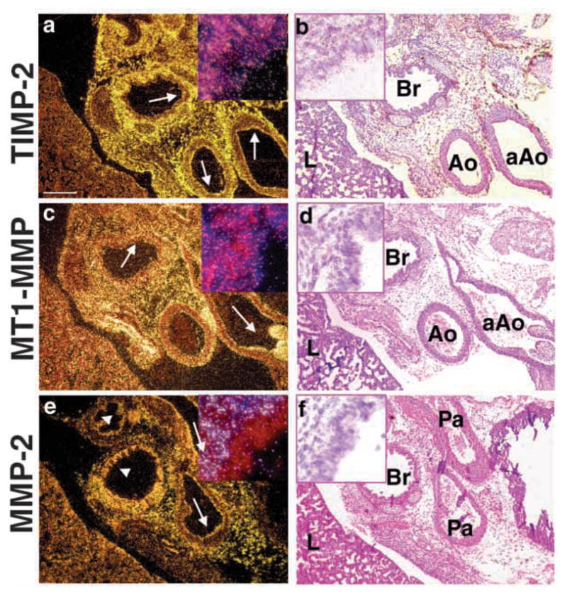
Localization of MMP-2 mRNA in the mesenchyme. Embryos from normal fetuses were taken at E18.5. For ISH analysis the following antisense probes were used: (a) TIMP-2, (c) MT1-MMP, (e) MMP-2. (b,d,f) H and E counterstain of darkfield images are shown. Note localization of TIMP-2 and MT1-MMP to the epithelia and mesenchyme (arrows), and absence of MMP-2 in the epithelia (arrowheads). Inset in each panel is 60× magnification of bronchial region from the corresponding image. aAo, ascending aorta; Ao, aorta; Br, bronchus; L, lung; Pa, pulmonary artery. Bar, 200 μm (a–f).