
Figure 1. Targeted expression of K-RasG12D to pancreatic acinar cells at endogenous levels induced non-progressing mPanIN-1 lesions unless p53 was deleted.
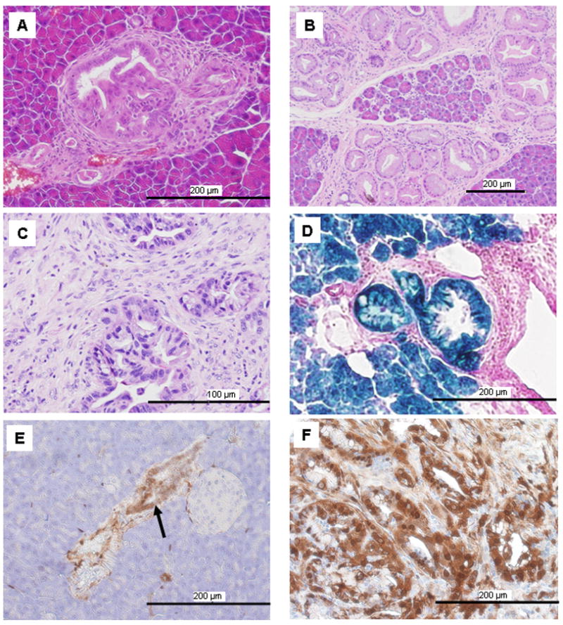
(A) mPanIN lesions developed in a 2-month-old LSL-KRas/Ela-CreERT mouse. (B) The number of mPanINs increased as the mice aged but did not progress (15 months old). (C) Mice (LSL-KRas/p53f/f/Ela-CreERT) which possessed endogenous expression of mutant K-Ras and p53 deletion readily developed PDAC (5 months old). (D). For lineage tracing, LSL-KRas/Ela-CreERT mice were further crossed with Rosa26 reporter mice such that cells of acinar lineage stained for β-galactosidase activity (blue). All mPanINs cells stained blue. (E). mPanIN cells (arrow) had elevated levels of p-Erk compared to surrounding acinar cells in a 4-month-old LSL-KRas/Ela-CreERT mouse detected using an anti-p-Erk antibody. (F). Cancer cells in PDAC developed from LSL-KRas/p53f/f/Ela-CreERT mice (5 months old) also showed strong p-Erk staining.