Figure 4.
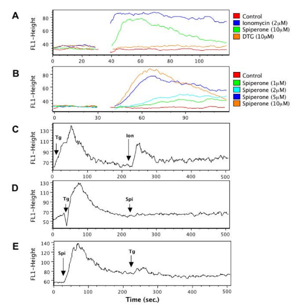
Spiperone induce a rise in intracellular calcium levels. (A) Tracing of fluo-4 fluorescence in cells stimulated with 10 μM spiperone, 10 μM DTG and 2 μM ionomycin over time. Drugs were added at 30 sec. (B) Tracing of fluo-4 fluorescence in cells stimulated with increasing doses of spiperone. Drugs were added at 30 sec. (C) Pretreatment with 1 μM thapsigargin could not abolish ionomycin-induced calcium release. Drugs were added at 10 and 225 sec as indicated by the arrowheads. (D) Thapsigargin blocks the subsequent cellular calcium responses to spiperone. Intracellular Ca2+ was recorded after stimulation with 1 μM thapsigargin and 10 μM spiperone. Drugs were added at 30 and 225 sec as indicated by the arrowheads. (E) Spiperone prevented thapsigargin-induced calcium increase. Intracellular calcium was traced after stimulation with 10 μM spiperone and 1 μM thapsigargin. Drugs were added at 30 and 225 sec as indicated by the arrowheads.

