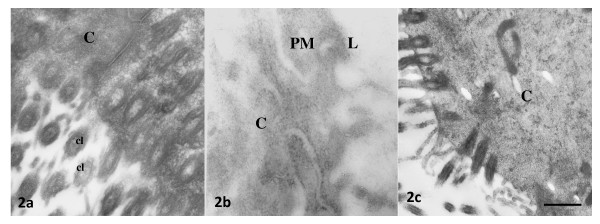
Figure 2.
Representative photomicrographs of oviductal epithelial cells processed by immunoelectron microscopy without prior incubation with anti-ESR1 antibody (a) or anti-ESR2 antibody (b), or incubated with rabbit preimmune serum (c). Bar: 0.5 μm. PM = plasma membrane, C = cytoplasm, L = lumen, cl = cilia. Bar: 0.5 μm.

