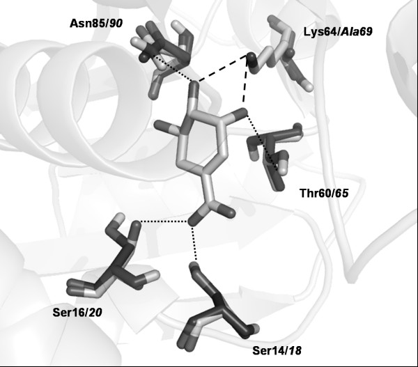
Figure 4.
MtbSD K69A model superimposed on experimentally solved T. thermophilus SD structure. Amino acid side chains involved in SHK binding and SHK molecule are shown as sticks. T. thermophilus and MtbSD K69A amino acids are colored, respectively, in light gray (residue number in bold) and dark gray (residue number in italics). H-bonds are shown as dotted lines; dashed lines represent H-bonds between Lys64/69, missing in MtbSD K69A model.

