
Figure 4. Recombinant HGF acting through c-Met signaling stimulates invasive outgrowths and increased secretion of uPA and uPAR from DCIS cells.
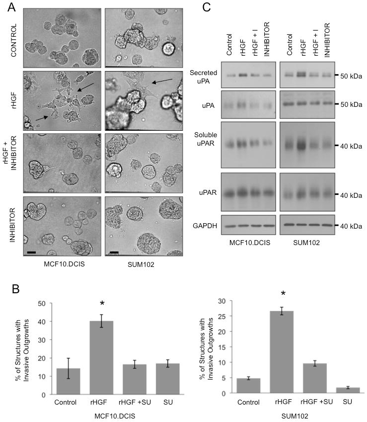
MCF10.DCIS and SUM102 cells were grown in 3D rBM culture with either 100 ng/ml HGF (rHGF), 100 ng/ml HGF + 2 μM SU11274 (rHGF + I), 2 μM SU11274 (I) or DMSO (Control). A, Representative DIC images depicting condition before harvesting cell lysates and conditioned media; bar, 200 μm. B, Images from MCF10.DCIS and SUM102 cell cultures were scored for the number of total structures and those with invasive outgrowths. C, Cell lysates and concentrated CM were separated by 12% SDS-PAGE under non-reducing conditions with lysates loaded based on protein concentration and CM loaded based on the protein concentration of the corresponding cell lysates. Proteins were transferred to nitrocellulose membranes and analyzed with antibodies against uPA or uPAR.