
Figure 5. HGF increased degradation of DQ-collagen IV by MCF10.DCIS and SUM102 3D cultures.
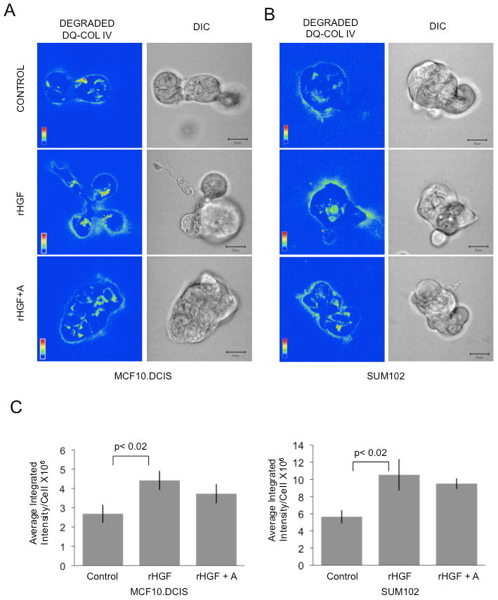
MCF10.DCIS or SUM102 cells grown in 3D rBM cultures containing DQ-collagen IV were treated with 100 ng/ml HGF (HGF), 100 ng/ml HGF plus aprotinin (HGF+A) or left untreated (Control). Integrated fluorescence due to proteolysis (green) was normalized to the number of cells (nuclei; blue). Representative fluorescence micrographs of one confocal plane of both the degraded DQ-collagen IV channel (degraded DQ-IV) and the corresponding DIC channel are illustrated for MCF10.DCIS (A) and SUM102 (B) 3D rBM cultures. Bar, 20 μm. C, Quantification of proteolysis in 3D structure volumes. Data were pooled from 3 representative experiments and are presented as mean ± SEM (n=18). Student's t-test, p<0.02.