
Figure 4. Immunohistochemical analysis of TAGLN, TAGLN2, and PPIA expression in pulmonary adenocarcinoma.
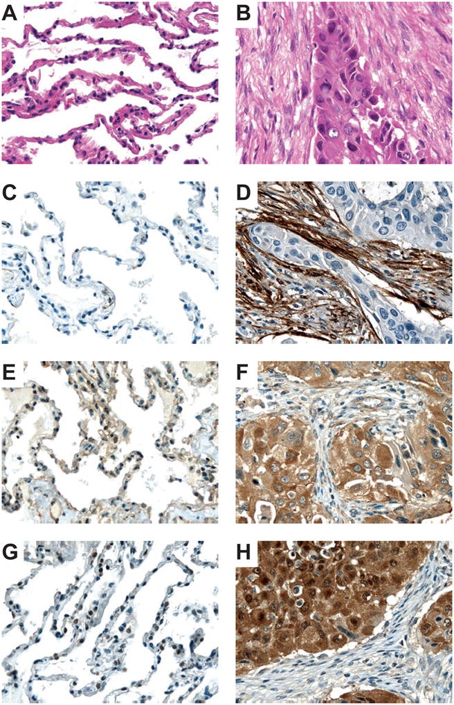
Photomicrographs of normal lung tissue (A) and lung adenocarcinoma (B) stained with hematoxylin-eosin. Protein expression of TAGLN (C, D), TAGLN2 (E, F), and PPIA (G, H) in lung tissue (left, normal; right, adenocarcinoma). Presence of the specific protein is indicated by the amount of brown staining. Nuclei were counterstained with hematoxylin (blue) for visualization purposes. The tissue shown is from patient LA13. Original magnifications: 400×.