
Figure 5. Immunohistochemical analysis TAGLN2 expression in atypical adenomatous hyperplasia (AAH).
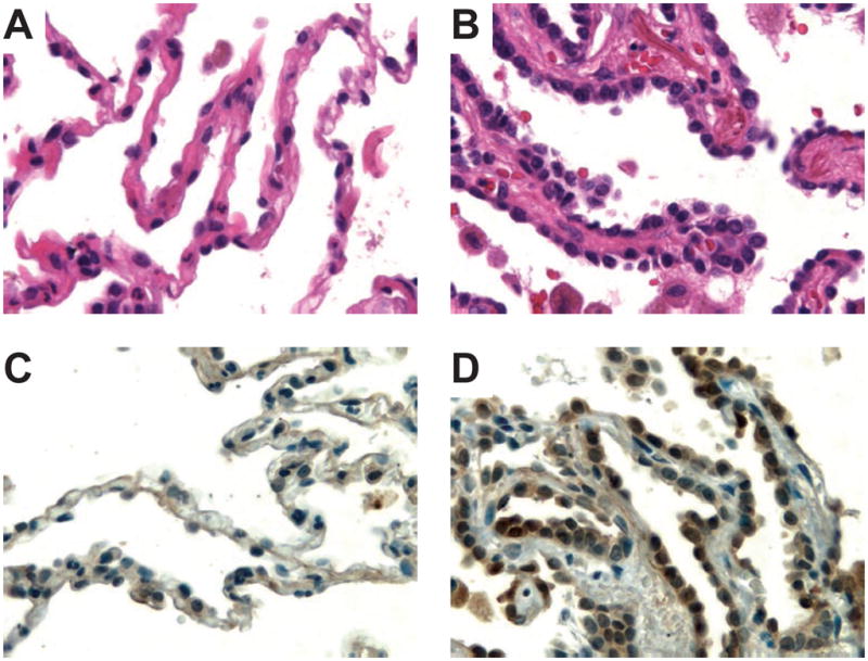
Photomicrographs of normal lung tissue (A) and a focus of AAH (B) stained with hematoxylin-eosin. Protein expression of TAGLN2 (C, D) in alveolar pneumocytes (left, normal; right, atypical adenomatous hyperplasia). Presence of the specific protein is indicated by the amount of brown staining. Nuclei were counterstained with hematoxylin (blue) for visualization purposes. Note the histopathologic features of AAH, such as slightly thickened alveolar walls and an increased number of alveolar lining cells with cytologic atypia. The tissue shown is from patient LA11. Original magnifications: 640×.