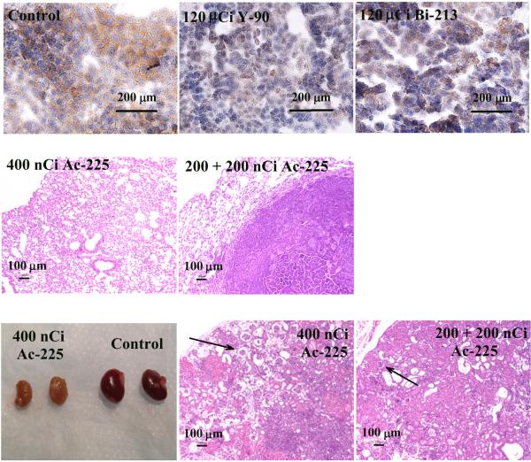
Figure 4.
A, Immunohistostaining of rat HER-2/neu expression on lung metastases. Untreated control (left); treated with 120 μCi 90Y-7.16.4, (middle) or with 120 μCi 213Bi-7.16.4 (right). B, H&E staining of lungs from mice treated with 400 nCi 225Ac-7.16.4 that showed no sign of tumor cells or lung tissue damage (left) and of a single metastasis found on the lungs of a mouse treated with 200 + 200nCi 225Ac-7.16.4 (right). C, left, Representative photographs of kidneys from a neu-N mouse surviving one year after treatment with 400 nCi 225Ac-7.16.4 (left pair) and a healthy neu-N mouse (right pair). H&E staining of kidneys from neu-N mice surviving one year after treatment with 400 nCi (middle) or 200 nCi + 200 nCi 225Ac-7.16.4 (right). Arrows point to collapse of cortical tissue due to loss of tubular epithelium in the kidney cortex. Bar indicates 100 μm.

