
Figure 1.
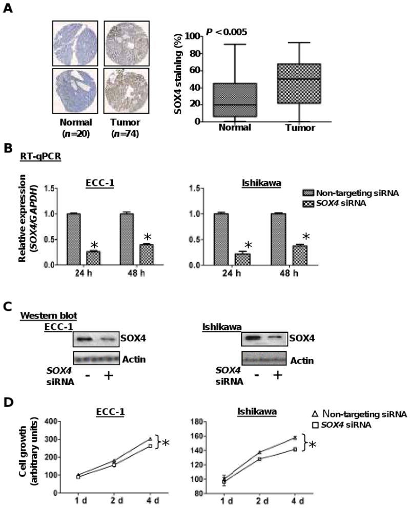
SOX4 is overexpressed in endometrial tumors. A, representative photographs of endometrial tissue microarrays (1.5mm core diameter) which underwent immunohisochemical staining for SOX4 and were scored for nuclear staining by the TissueMine software. Boxplots of SOX4 nuclear staining for normal tissue (n=20) and endometrial tumors (n=74) P < 0.005. B & C, relative expression levels of SOX4 mRNA and protein in ECC-1 and Ishikawa cells after transient transfection with SOX4 siRNA or a pool of non-targeting siRNA oligonucleotides for 24 and/or 48 h. GADPH and β-actin served as internal controls for RT-qPCR and western blotting respectively. Error bars represent SD among triplicates; *, P<0.05. D, cellular proliferation was measured by MTS assay in endometrial cancer cells transfected with SOX4 siRNA or non-targeting siRNA. Transfectants (3,000/well) were placed in 96-well plates and proliferation was measured every 24 h. Each point represents the mean value of at least triplicates. *, P < 0.05.